

Feline Leukemia Virus (FeLV) is a virus belonging to the genus Gammaretrovirus within the family Retroviridae. It is transmitted through sources such as saliva, feces, and shared food and water bowls. FeLV can cause various hematopoietic tumors, particularly lymphoma.
Virology
Feline Leukemia Virus is a positive-sense, single-stranded RNA virus classified within the genus Gammaretrovirus of the family Retroviridae. It possesses the enzyme reverse transcriptase, which enables the synthesis of DNA from RNA.
Among the envelope proteins, p15E is considered to have immunosuppressive properties, while gp70 is clinically significant because it determines the viral subgroups (A, B, C, etc.). These subgroups may occur individually or in combination within the same cat. Envelope proteins constitute important components of FeLV vaccines.
Among the core proteins, p27 is of particular importance, as it is detected by ELISA and IFA tests and is widely used in the diagnosis of FeLV infection.

FeLV Subtypes
FeLV subtypes can be detected in various combinations; however, subtype A is the only naturally transmissible form. Significant antigenic variations exist among subtypes. Although FeLV-A alone exhibits limited pathogenicity, its combination with other subtypes markedly increases disease severity.
Cats infected with FeLV-A and FeLV-B commonly develop thymic lymphoma. In contrast, FeLV-C is associated with erythroid hypoplasia and severe anemia, often resulting in death within 1–2 months. It is also known that recombination with proto-oncogenes can lead to the formation of additional subgroups.
Epidemiology
FeLV is distributed worldwide and affects both domestic and wild felids. The implementation of testing and quarantine protocols, vaccination programs, and exclusion of infected animals from breeding has significantly reduced its prevalence. The infection is more commonly observed in young cats and in environments with high population density, such as multi-cat households or shelters, particularly where hygiene conditions are inadequate.
Transmission
The virus is transmitted between cats via virus-containing secretions. Close contact and shared food and water bowls represent significant sources of infection. During the viremic phase, high viral loads are detected in saliva. The virus has also been identified in feces, urine, and milk. Transmission via these secretions may occur indirectly as well.
FeLV can remain infectious for up to 48 hours at 37°C and for 1–2 weeks at 22°C in blood or secretions. However, because the virus is sensitive to heat and most disinfectants, the risk of transmission from regularly cleaned surfaces such as veterinary examination tables is very low. In contrast, facilities with high animal turnover and shared environments, such as cat boarding facilities, pose a higher risk if adequate disinfection is not maintained.
Pathogenesis
The virus enters the body via the oral and nasal routes (oro-nasal transmission). After entry, it initiates infection by infecting lymphocytes in the oropharyngeal region. This is followed by primary viremia, during which infected lymphocytes disseminate throughout the body.
The virus subsequently localizes in the bone marrow and infects hematopoietic cells. This leads to secondary viremia, after which the virus colonizes epithelial cells, particularly in the salivary glands and intestinal mucosa, resulting in viral shedding through saliva and feces.
Cats that clear viremia are considered latently infected. In these cats, FeLV may be reactivated by corticosteroid use or stress. Cats that successfully overcome infection may develop long-lasting immunity.

Clinical Signs
The genetic basis of tumors induced by FeLV and its recombinant forms has not yet been fully elucidated. However, FeLV is known to cause a wide range of hematopoietic neoplasms, among which lymphoma is the most common tumor in cats. Clinical manifestations vary depending on tumor localization. In some cases, tumors may be associated with another virus, Feline Sarcoma Virus (FeSV).
The prognosis in persistently viremic cats is generally poor. Although not all lymphomas are FeLV-associated, FeLV-positive cats have a 62-fold increased risk of developing lymphoma compared to FeLV-negative cats.
Diagnosis
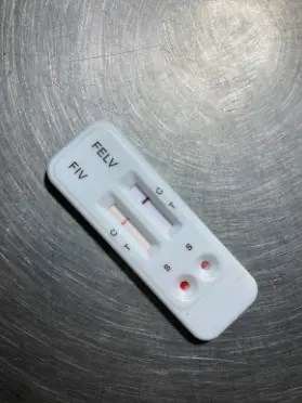
Cats belonging to risk groups should be tested for FeLV. These include cats showing clinical signs, suspected cases, shelter cats, and cats with outdoor access. Antigen detection can be performed using ELISA and IFA tests. Detection of antibodies by ELISA is not preferred, as it depends on the stage of viremia.
For definitive diagnosis, PCR testing may also be performed. Since approximately 10% of PCR-positive cats may test negative by ELISA, PCR confirmation is considered beneficial.
Prevention and Control
Cats infected with FeLV should be protected from exposure to other infectious agents; therefore, infected animals must be kept under quarantine. In multi-cat households, all cats should be tested, and infected individuals should be isolated. Cats may be reintroduced to the group only after two consecutive negative FeLV test results.
The same quarantine and testing procedures apply to newly introduced cats. Additionally, FeLV-positive cats should undergo clinical and serological evaluations twice annually.
Vaccination
The first FeLV vaccine was introduced in 1985, and numerous vaccines have since become available. Cats should be tested for FeLV prior to initial vaccination. Vaccination primarily aims to prevent viremia and is generally recommended for cats at higher risk of exposure. Routine vaccination is not typically advised for neutered, strictly indoor cats.
In kittens, vaccination is administered as two doses at 8 and 12 weeks of age, followed by a single booster one year later. Subsequent booster vaccinations are determined based on the individual cat’s risk category.
This blog post has been verified by DVM Elif KENAR.
Resources
- Vail, D. M., Thamm, D. H., Lipták, J. M. (2020). Withrow & Macewen’s small animal clinical oncology. Elsevier.


